Root coverage in Jeju: restoring receding gums around a crowned canine
The old crown was removed and replaced with a temporary crown before gum grafting and suturing. This record follows early healing and placement of the final crown.
Treatment at a glance
- Initial findings
- Gum recession had exposed the root and old crown margin of an upper canine.
- Treatment plan
- The crown was removed, and root coverage was performed while a temporary crown was in place.
- Treatment
- Gum grafting and suturing were followed by placement of the final crown.
- Early follow-up
- Photographs document the site one week after surgery and the final crown at about three weeks. Gum healing and maturation were still continuing.
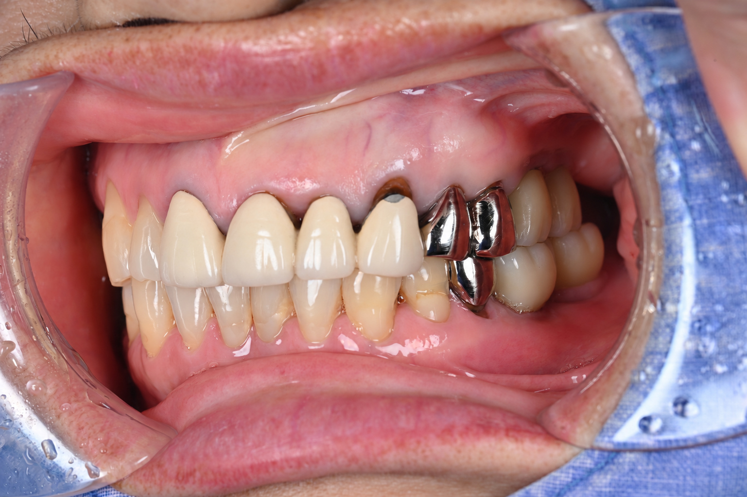
The root and old crown margin were exposed
The gum around an upper canine had receded, exposing the tooth root and the margin of the existing crown.
The photograph records the relationship between the old crown and the surrounding gums before treatment.
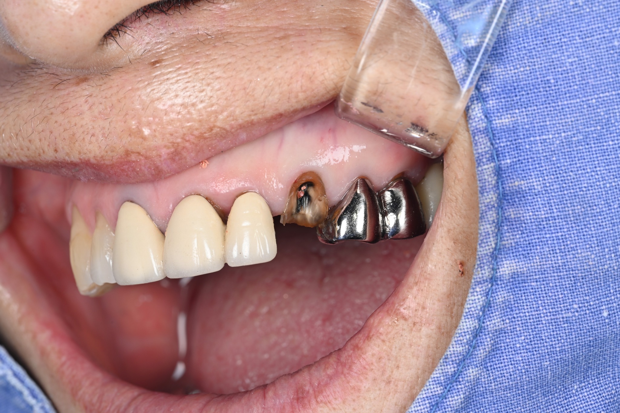
Removing the old crown and placing a temporary crown
The existing crown was removed. The photograph shows the tooth and surrounding gums after crown removal.
A temporary crown was then fitted. Gum grafting was performed with this temporary crown in place.
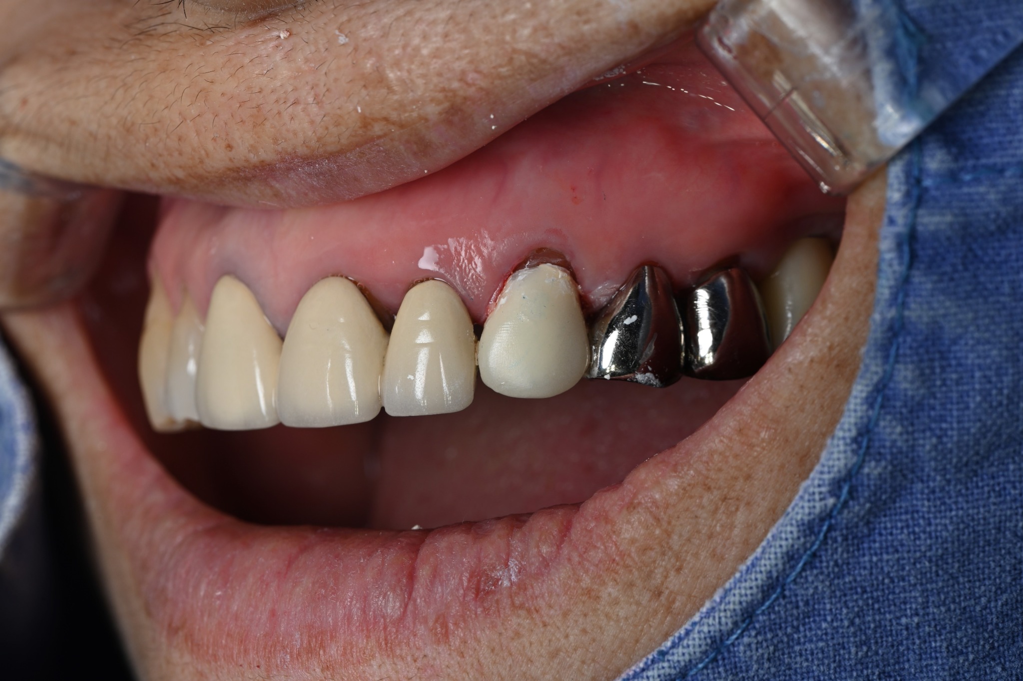
Gum grafting compared with healing at one week
Gum grafting was performed at the exposed root. The tissue was repositioned to cover the root and secured with sutures.
The temporary crown remained in place at this stage; the final crown had not yet been fitted.
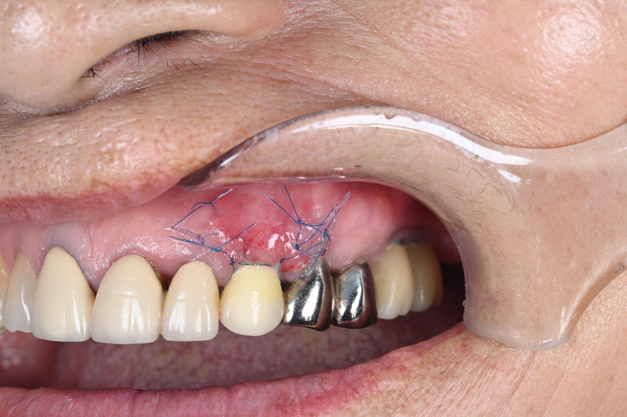 1 week after surgery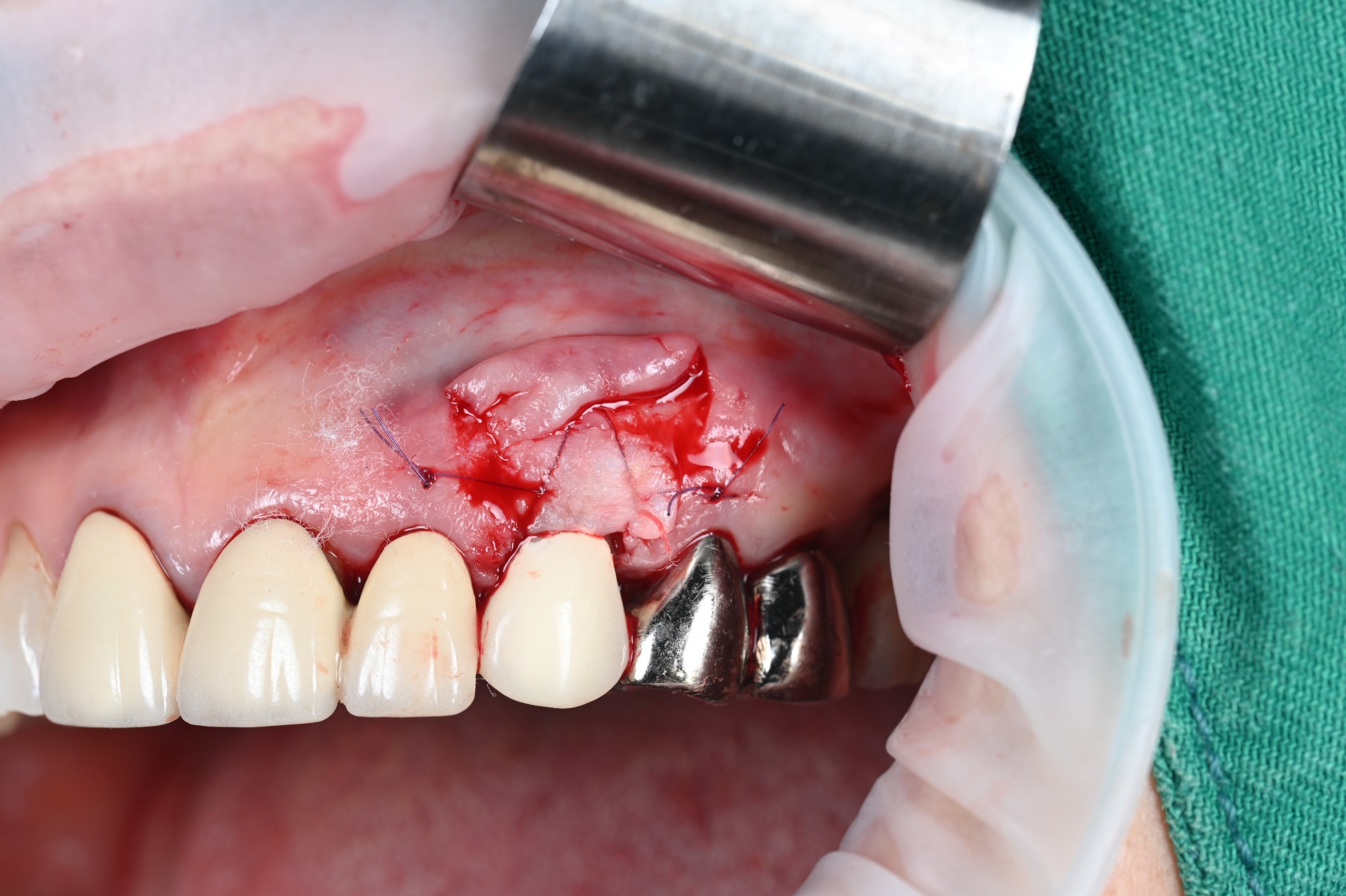
One week after surgery
The photograph was taken one week after surgery. Sutures remained, and the canine still had its temporary crown.
This is an early healing record. The shape and colour of the gums may continue to change as healing progresses.
Completing treatment with the final crown
The temporary crown was later replaced with the final crown. These photographs record the final crown at about three weeks after gum surgery.
Compared with the photograph before treatment, the previously exposed root above the canine is covered by gum tissue, and the appearance where the new crown meets the gums has changed.
Treatment progressed from old crown removal and temporary crown placement to gum grafting and final crown placement.
Gum healing and maturation continue after the crown is fitted. These photographs document early progress; long-term stability is assessed at subsequent regular reviews.
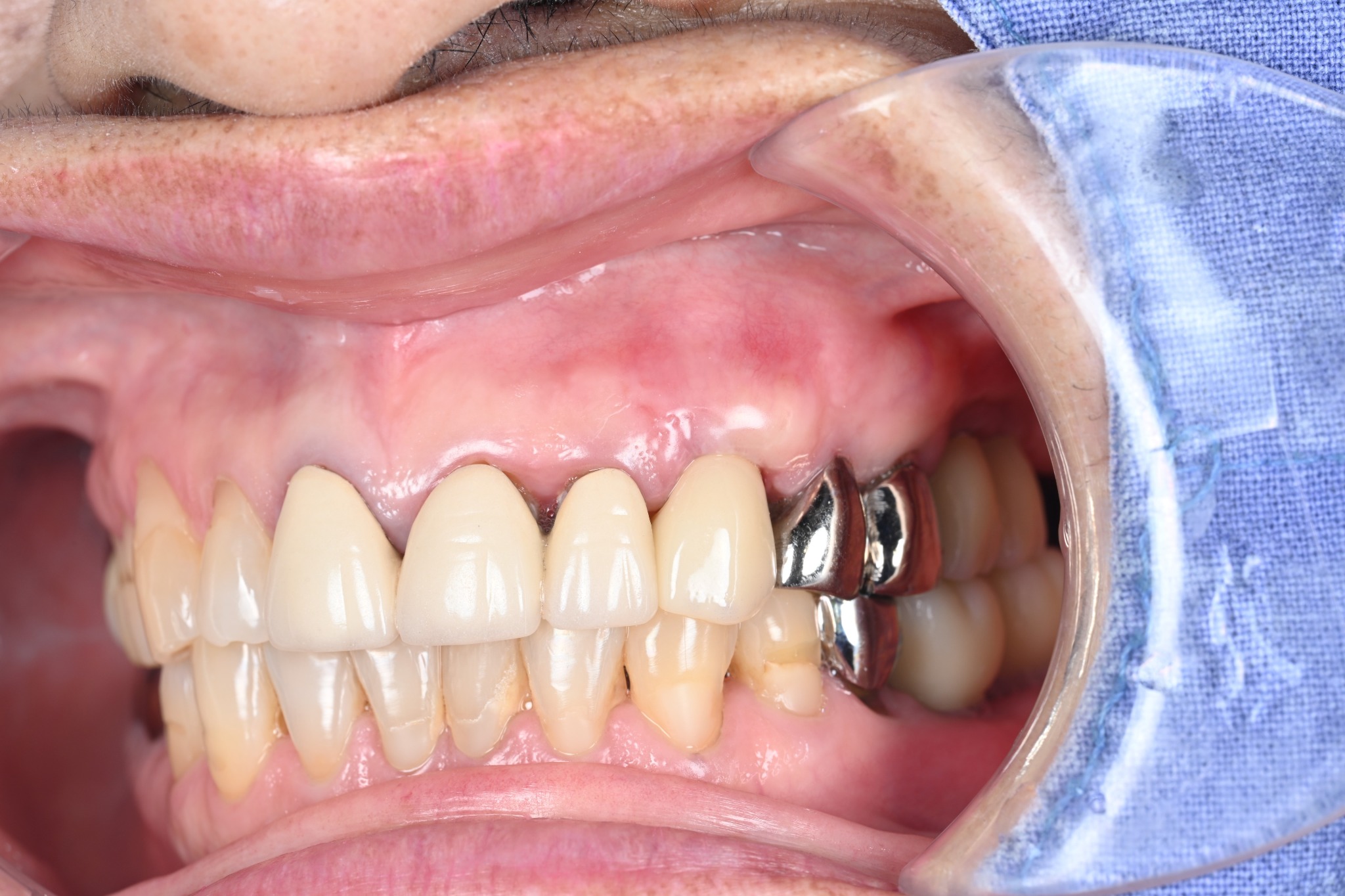 About 3 weeks after surgery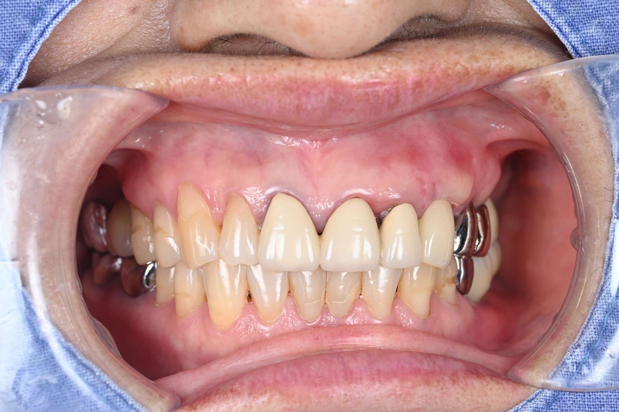
Read more
This clinical journal describes an actual treatment case with identifying information excluded.
Treatment methods, duration, and outcomes may vary according to general health, oral condition, bone, bite, and maintenance needs.
This page provides general medical information and does not replace an individual examination or diagnosis.