Dental implants in Seogwipo: how do diagnosis, planning and the final teeth connect?
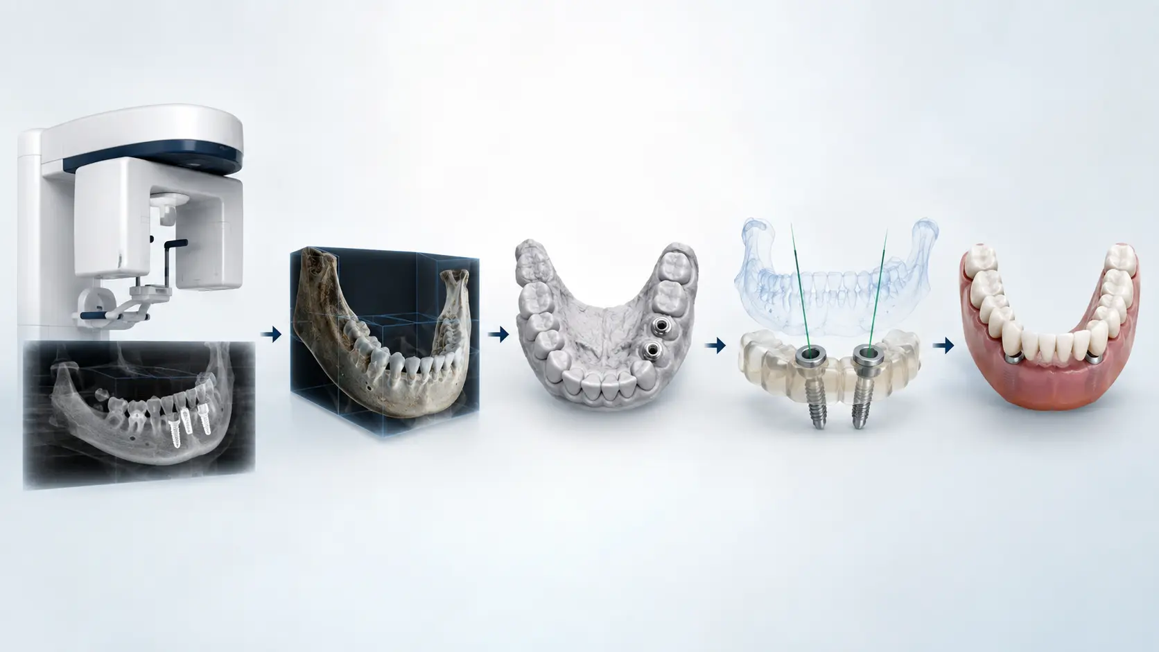
Bone quantity, implant number and operation time are useful questions, but the aim of treatment is comfortable, maintainable teeth.
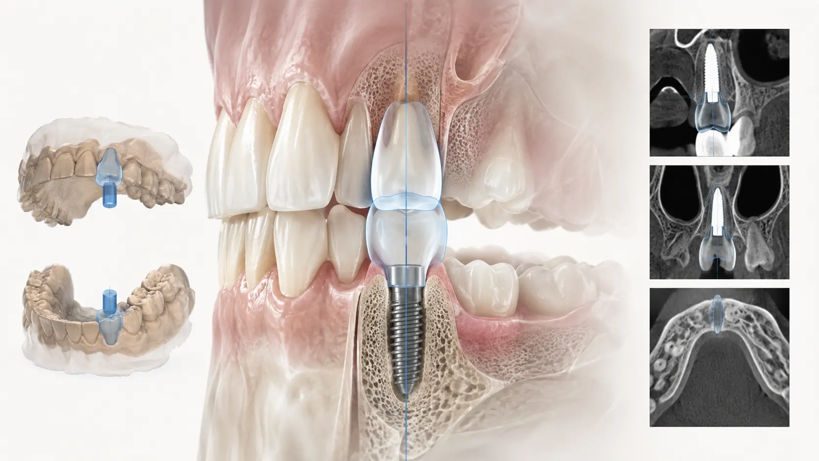
The position and shape of the final teeth should guide the implants supporting them. Diagnosis, surgery, temporary teeth and final teeth therefore need one coordinated plan.
Key points
Work back from the intended final teeth, check the plan against the actual mouth and carry lessons from temporary teeth into the final restoration.
Why plan backwards from the final teeth?
Even a firmly integrated implant can leave an awkward restoration if it is too far inside or outside the intended tooth position or has an unfavourable screw opening.
Food traps, inaccessible cleaning spaces and concentrated bite forces can stress the restoration, screws and surrounding tissues. Planning seeks a position that satisfies both tooth position and biological conditions.
- Define the shape and position of the missing teeth
- Check opposing teeth, smile, speech and cleaning space
- Plan implant depth, direction and diameter to support those teeth
- Confirm that bone and anatomical structures permit the plan
How do CT and oral scans complement each other?
CT shows bone dimensions and shape, nerves and sinuses. It does not capture the tooth and gum surfaces or bite as precisely as an oral scan.
The oral scan records these surfaces and spaces. Aligning the two datasets allows assessment of final teeth, neighbouring structures, guide support and the bite.
The clinician must check alignment, movement or metal distortion and agreement with the mouth. More data does not automatically complete the diagnosis.
What is checked when transferring the plan to surgery?
A surgical guide can transfer planned direction and depth. Research reports that computer-assisted methods can improve reproduction of planned positions compared with freehand placement.
Deviation can still arise from guide fit or fixation, mouth opening, drill length, moving soft tissues and bone quality. A guide supports clinical judgement.
During surgery the clinician checks stable seating, achievable direction and depth, unexpected findings and whether initial stability supports the temporary-restoration plan.
Are temporary teeth only for the waiting period?
Temporary teeth are an assessment stage, especially for front teeth or connected implant restorations.
They help check length and width against the face and lips, speech, comfort, force distribution and practical cleaning. These findings should inform the final design. Changing material without correcting a problem can preserve the same discomfort.
What is checked in the final restoration?
Appearance is one part of the assessment. Connection stability, force distribution and cleaning access also matter.
At Hyungje Dental Clinic, oral scans, digital design and in-house fabrication help carry the shape and bite assessed in temporary teeth into the final restoration.
- Fit at the implant connection
- Contacts with neighbouring teeth
- Excessive contact when biting or moving sideways
- Cleaning space between gums and restoration
- Screw-opening position and material thickness
- Use of a toothbrush, floss and interdental brushes
Can digital equipment guarantee the result?
CT, oral scanners, software and guides cannot guarantee an outcome. Small deviations can accumulate through imaging, alignment, design, guide production, surgery, scanning, fabrication and fitting.
Healing, hygiene, smoking, medical conditions and grinding are also beyond the equipment alone. What matters is interpretation, checking differences between plan and reality and using temporary-restoration findings in final care.
What should the explanation before treatment include?
The patient should understand how the records inform design, surgery, temporary teeth, final teeth and maintenance. Hyungje Dental Clinic in Seogwipo aims to connect these stages consistently.
- Were the final teeth planned first?
- What did CT and oral scans show?
- Why were the implant position, angle and depth chosen?
- Why use a guide, and what are its limits?
- What will the temporary teeth be used to assess?
- How are the final material and attachment chosen?
- How will cleaning and reviews continue?
Related articles
References
- Wismeijer D, et al. Group 5 ITI Consensus Report: Digital technologies. Clin Oral Implants Res. 2018;29(Suppl 16):436–442.
- Khaohoen A, et al. Accuracy of implant placement with computer-aided static, dynamic, and robot-assisted surgery: a systematic review and meta-analysis of clinical trials. BMC Oral Health. 2024;24:359.
- Reiff FS, et al. Accuracy of Freehand, Static, and Dynamic Computer-Assisted Implant Placement: A Systematic Review and Meta-Analysis. J Periodontal Res. 2026;61(2):111–137.
This article provides general dental information. Individual diagnosis and treatment require an examination.